Table of Contents
What is the Cornea?Diseases & ConditionsTypes of Corneal SurgeryEye Banking 101Financing Corneal Procedures in Overland Park, KS
Dr. Cavanaugh has the training, experience, and unsurpassed technology to provide you with optimal corneal surgery results
Dr. Cavanaugh at Cavanaugh Eye Center is one of the area’s leading laser eye surgeons. As a fellowship-trained cornea specialist, he is knowledgeable of all types of corneal conditions and able to perform a wide array of corneal surgeries for patients whose corneas have been damaged due to disease, trauma, or heredity. Contact Cavanaugh Eye Center to find out how we can help improve your vision. We look forward to seeing you for your comprehensive consultation for corneal procedures in Overland Park, KS.
Schedule a Consultation Today!
What is the Cornea?
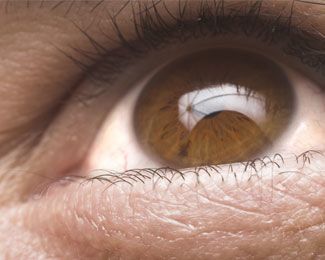
The cornea is the clear dome-shaped outer covering of the eye over the iris (the colored part) and the pupil. It bends, or refracts, light rays as they enter the eye, and it is often referred to as the “window of the eye.” The cornea acts as the eye’s first curved focusing structure, which holds two-thirds of the eye’s focusing power. Since light is first passed through the cornea, it is necessary to have both a clear and smooth cornea to allow it to focus incoming light rays precisely on the retina at the back of the eye so you can see clearly. The cornea has five main layers and disease can occur in any or all of the five layers.
From the outside to the inside, the cornea’s layers include:
- Epithelium
- Bowman’s Membrane
- Stroma
- Descemet’s Membrane
- Endothelium
Corneal Diseases & Conditions
If the cornea becomes cloudy, swollen and/or scarred, your vision will be blurred or reduced. This occurs as a result of injury, degeneration, infection, or inherited dystrophy. Examples of such conditions are:
- Hereditary corneal dystrophies: Fuchs’, Lattice, Granular, Macular
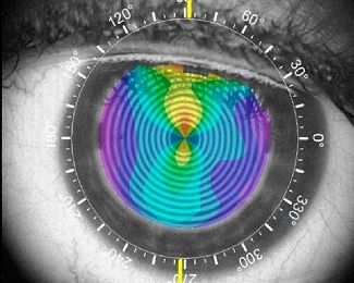
- Keratoconus: cone-like steepening of the cornea
- Scarring after infections: herpes virus, bacterial, fungal, etc.
- Scarring after injury
- Corneal failure after other eye surgery such as cataract surgery
- Rejection after corneal transplant
Types of Corneal Procedures
Treatment is tailored to the individual disease and the individual patient. The underlying disease and contributing problems need to be addressed. Depending on the condition, corneal procedures might include medications, laser treatment, corneal transplantation, or other surgery. Treatment modalities are often determined by which layer(s) of the cornea are involved.
CXL is used to halt the progression of Keratoconus and prevent the need for more invasive treatment options. CXL utilizes a vitamin B2 solution and UV light applied to the cornea to help strengthen the corneal collagen fibers by cross-linking them together.
Penetrating Keratoplasty (PK) is a full-thickness corneal transplant and is best for conditions that involve clouding throughout the entire cornea or for conditions such as keratoconus where the cornea’s shape is severely distorted. In this surgery, all five layers of the cornea are removed and replaced with a clear donor cornea.
DSAEK replaces the posterior or inner layers of the cornea and is used for selected patients with Fuch’s dystrophy or other disorders involving the endothelial or inner layer only.
Phototherapeutic Keratectomy (PTK) is a procedure that uses an excimer laser to remove haziness and irregularities from the cornea.
Superficial Keratectomy (SK) removes scars and/or defective surface cells from the cornea so that new healthy cells can take their place.
Amniotic membranes are rich with fetal stem cells and contain growth factors that promote wound healing on the surface of the eye. Amniotic membrane therapy has been found to be a good alternative for corneal and conjunctival reconstruction in many clinical situations.
PROKERA is a medical device used to protect, repair, and heal damaged eye surfaces that are made by setting a piece of amniotic membrane tissue between two rings made out of a clear, flexible material.
Eye Banking 101: How We Obtain your Donor Cornea
In the Midwest, we are fortunate to have an excellent eye bank system. The Saving Sight eye bank supplies corneal tissue to many transplant recipients in the Midwest. As a past Medical Director of the Kansas City Eye Bank, Dr. Cavanaugh was instrumental in helping forge what is now the Saving Sight organization. Because of this network and our close relationship with Saving Sight, we are now able to schedule corneal transplants like any other surgery and there is generally no waiting list.
The Kansas City branch of Saving Sight is the headquarters and functions as the DSAEK tissue processing center for the entire Midwest. In this facility, the donor cornea is precisely split so that only the posterior (back) layers can be transplanted. Dr. Cavanaugh was asked to oversee the training and certification of the technicians and the new state-of-the-art tissue preparation facility. View Dr. Cavanaugh’s recognition from Saving Sight.
Your donor cornea is received through Saving Sight from a person who has recently passed away. This person’s family has decided to allow their loved one to continue living by donating usable tissue. You may receive a cornea from a local donor or an out-of-state donor. All tissue is tested for the HIV/AIDS virus, and Hepatitis B and C. Strict screening criteria must be met before tissue qualifies for transplantation. When the eye bank receives the corneas, they call the doctors who have patients scheduled for surgery within the next few days and notify them of tissue availability. If Dr. Cavanaugh
is unable to acquire suitable tissue for you, one of his staff will call you and reschedule your surgery for later. This occurrence is very uncommon.

Financing Your Cornea Transplant Surgery
Many corneal treatment options and procedures are covered due to their medical nature, however, non-covered services are the patient’s responsibility. Your unique health insurance plan dictates your deductible amount, copayments, or other co-insurance. Our skilled billing department will do its best to provide you with an accurate estimate of benefits and out-of-pocket expenses. More information is available here.
We require 24-hour notice of rescheduled/canceled appointments to avoid a fee.
If you have health insurance coverage:
- Please supply us with a current copy of your insurance card.
- Please notify us of any changes in your address or telephone number.
- All copays are due at the time of service.
- All non-covered services such as refractions (new prescription for glasses) and elective procedures will be due at the time of service.
- All referrals are the responsibility of the member/patient and must be current prior to your visit.
- Your estimated portion, including any deductibles, co-insurance or non-covered service will be expected to be paid prior to your surgery date (our business office will notify you in advance if this is required).
- Patient balances are due 30 days after the insurance notifies our office of patient responsibility.
- Patient will be responsible for any fees associated with enforcement of collection action.
If you do not have health insurance, payment in full is required at or before the time of service.
As a courtesy, we will gladly assist you by filing your claim with your insurance carrier. If you receive a statement from our office after insurance has paid or denied, the balance then becomes your responsibility to pay. If you disagree with the balance for any reason please contact our business office at 913-752-9675.
We accept cash, check, Visa, MasterCard, Discover, and American Express. We do not provide financing in-house for cornea surgery however financing arrangements can be made through CareCredit. The CareCredit process is quick and efficient.
CareCredit offers a full range of payment plans so you can find one that works best for you. With the popular ‘Deferred Interest’ plans, there are no interest charges if you pay your balance in full within the specified time period. CareCredit offers 12, 18, or 24-month plan options with low monthly payments available. You apply via https://www.carecredit.com/go/FWJ652/ and your specific rates will depend on your credit rating. The process is simple and you will have a credit determination quickly. Our surgical counselors can provide you information about CareCredit and help guide you through the process.
Contact Us for Corneal Procedures in Overland Park, KS
At Cavanaugh Eye Center, we are dedicated to providing patients with the best vision care and can help answer all of your questions about corneal procedures in Overland Park, KS. We look forward to meeting you at your consultation and thank you in advance for trusting your surgical care to us!
Are you ready to discuss your cornea surgery options?